Cholesteatoma

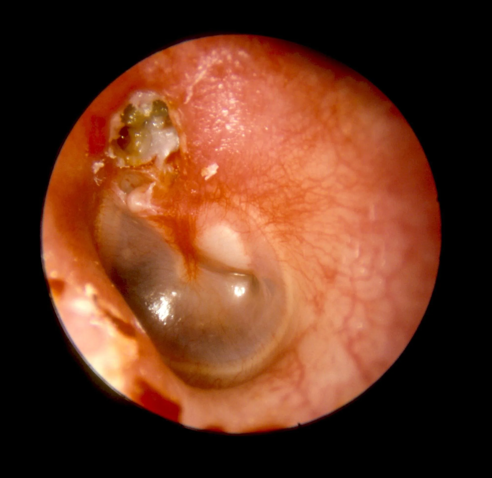
The photo on the left shows a normal ear canal, while the photo on the right is an ear drum with a cholesteatoma.
The outer surface of the ear drum is lined with skin, which (under normal circumstances) sheds off and comes out of the ear in wax. In a cholesteatoma, an area of ear drum becomes sucked in, creating a ‘retraction pocket’. The pocket slowly becomes deeper and deeper, and eventually gets to a point where the shed skin becomes trapped and starts to build up into a skin cyst. You can see the crusty area at the top of the ear drum at 12 o’clock, which is the opening of the retraction pocket, and the pearly white bulge behind the drum at about 2 o’clock, which is the skin cyst within the retraction pocket.
Although it sounds fairly harmless, these skin cysts can cause a lot of problems if left untreated. Because they are slowly expanding in a confined space, they can erode away at structures within the ear (ossicles/hearing bones, inner ear hearing and balance, facial nerve), but also can case infections which occasionally may be serious or life-threatening.
Symptoms
Symptoms caused by destruction of structures within the ear:
hearing loss
dizziness
facial weakness
Symptoms caused by infection:
smelly, discharging ear
mastoiditis (infection in the bone behind the ear)
meningitis/brain abscess
The commonest symptoms are hearing loss and persistent or recurrent snotty discharge from the ear that doesn’t clear up despite repeated courses of treatment. Meningitis, brain abscesses and facial weakness are thankfully rare.
Treatment
In most people with cholesteatoma, surgery is the only treatment once it has become established. There are various different surgeries (e.g. modified radical mastoidectomy, combined approach tympanoplasty), and the choice depends on several different factors (e.g. your anatomy, the size of the cholesteatoma, other health considerations). The general principles of cholesteatoma surgery is to remove the disease to leave an ear that is safe and dry, and to minimise the chances of recurrence. If hearing has been destroyed or damaged by the skin cyst it is sometimes possible to restore this, depending on the configuration of hearing bones (ossicles) that remain. However, this may need to be done at a second operation a year or so down the line, and it may only be possible to restore part of the hearing (or none at all). Regardless of the surgical technique used there is a risk of recurrence and ongoing surveillance is required for several years after surgery, either with an MRI scan or a second-look operation.
Risks
Bleeding, infection, recurrence, need for further surgery, taste disturbance, hearing loss, tinnitus, dizziness, facial weakness, ear asymmetry, conversion to mastoid cavity, need for lifelong ear cleaning in clinic.